
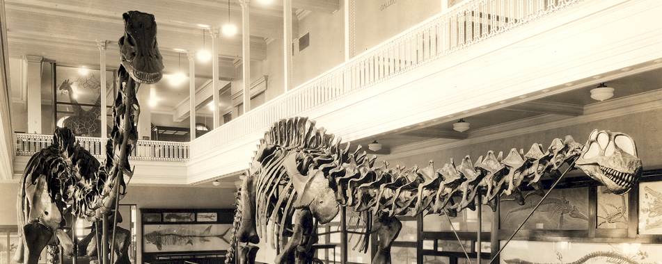
Key Points
- Osteology is the study of the skeleton.
- The bodies of vertebrates have homologous structures (the same body part, regardless of its particular shape and function) as well as analogous structures (non-homologous parts used for the same function).
- Bones and other features of anatomy are given formal names (normally based on Latin).
- Anatomical directions and landmarks are used to described the position of different anatomical features.
- Major sections of the skeleton include the skull, the axial skeleton (vertebral column and other parts along the main trunk of the body), and the appendicular skeleton (the pectoral girdle and forelimb, and the pelvic girdle and hindlimb.)
Introduction to Anatomy

Labeled skeletons of Camarasaurus and Tyrannosaurus; image courtesy of Sauropod Vertebra Picture of the Week
Homologous structures: the same anatomical structure, regardless of function.
Analogous structures: represent different units of anatomy serving the same function.
Comparative anatomy seeks to describe the structure of the bodies of organisms in terms of their homologous structures.
Functions of the skeleton:
- Support
- Protection of organs: such as the facial bones protect the sensory capsules, the braincase protects the brain, the ribs protect the heart and lungs, etc.
- Mechanical Structures:
- Bones contact one another at joints. These joints may be immobile (like many joints in the skull) or permit motion (such as between vertebrae, in the jaws, or in the limbs)
- Bones are connected to other bones by ligaments; bones are connected to muscles by tendons
- Muscles pull by becoming shorter; they cannot PUSH (instead, a separate set of muscles has to act in order to move the joint back to the original position)
Anatomical Directions
Anatomical directions are used to describe the positions of anatomical structures relative to each other. These are intrinsic to the organism, and makes no reference to the position the animal is in nor its orientation in space. Anatomical directions come in complimentary pairs.

- Anterior (towards the tip of the snout)/Posterior (towards the tip of the tail)
- Dorsal (up and out through the spine)/Ventral (down and out through the belly)
- Medial (towards the middle)/Lateral (towards the sides)
- Proximal (towards the trunk)/Distal (away from the trunk)
- Proximal and distal are normally used only for the limbs, and occasionally for the tail
- (NOTE: some use "cranial/caudal" instead of "anterior/posterior" (and "rostral/caudal" within the head itself))
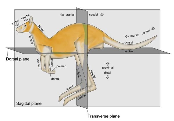
Anatomical views: when a specimen is illustrated, the anatomical view represents that surface of the specimen that is shown.
Anatomical landmarks: particular homologous structures on the skeleton (openings, joints, etc.) used for identifying the position of bones or other features of the anatomy.
The skeleton of a dinosaur (or other vertebrate) is divided into a couple of different sections:
- The skull, composed of:
- The cranium (braincase, face, and upper jaw)
- The mandible (lower jaw)
- The postcranium (everything posterior to the cranium), composed of:
- The axial skeleton (spine, ribs, and related features of the neck, trunk, and tail)
- The appendicular skeleton (forelimb, hindlimb, and their girdles)
(Incidentally, anatomical terms are for the most part based on Latin words. Bones or landmarks with Latin rather than English plurals are noted below)
Important Bones and Landmarks of the Skull
Important Bones and Landmarks of the Skull. NOTE: Almost all bones and landmarks of the skull are paired, with one on the right side and one on the left. Although the skulls of vertebrates are composed of many bones, these bones are joined by sutures: depending on the type of suture, the joint can be mobile or immobile.
- Orbit: eyesocket
- Naris (pl. nares): opening for the nasal passage. (Not exactly the same thing as the nostril, which is a fleshy structure.)
- Antorbital fenestra: a large opening in the facial bones of dinosaurs and their relatives, anterior to the orbit and posterior to the naris (fenestra, pl. fenestrae: a large opening in the skeleton, from the Latin word for "window")
- Teeth. In dinosaurs and most other land-dwelling vertebrates, the teeth are found in three main bones: two on each side of the upper jaw, and each side of the lower jaw
- Made of the same material (hydroxylapatite) as bone, but of hard dentine and even harder (more crystalline) enamel
- Dinosaur teeth (like mammals) grow out of "sockets" (alveoli)
- Like most vertebrates (but NOT mammals), they got new teeth from each socket throughout their life
- Socketed teeth consist of:
- A root (formed of dentine) with fits into the socket
- An enamel-covered crown that sticks out of the gumline
- In most dinosaurs, there is no tooth-to-tooth contact (occlusion): instead, the teeth swipe past each other
- The edges of many dinosaur teeth have either serrations (small lumps good for slicing meat) or larger denticles (large projections better for chopping up plants)
- Premaxilla (pl. premaxillae): anterior of the tooth-bearing bones of the cranium
- Maxilla (pl. maxillae): posterior of the two tooth-bearing bones of the cranium
- Lacrimal: bone separating the antorbital fenestra and orbit, contains the tear duct
- Postorbital: bone posterior to the orbit
- Jugal: the "cheek bone", ventral to the orbit
- Temporal fenestrae: openings in the back part of the skull for attachment and expansion of the jaw muscles. In dinosaurs and their relatives, there are two temporal fenestrae on each half of the skull (left and right):
- Infratemporal fenestra: also called the lateral temporal fenestra, opening on the side of the skull
- Supratemporal fenestra: opening on the top of the skull
- Nasal: bone along the top of the snout dorsal to the naris and maxilla
- Braincase: a collection of bones which surrounds the brain cavity
- Foramen magnum: Latin for "great opening", the hole in the back of the braincase where the spinal cord emerges from the brainstem
- Occipital condyle: a condyle (rounded knob joint) composed of several different bones just ventral to the foramen magnum; the connection between the cranium and the backbone
- Dentary: the tooth-bearing bone of the mandible; in mammals the whole of the mandible is composed of just the dentary, but in dinosaurs and most other vertebrates there are various postdentary bones
- Mandibular fenestra: in dinosaur and their relatives, an opening on the lateral surface of the mandible surrounded by the dentary and the postdentary bones
Teeth are composed of materials (softer dentine and harder enamel) similar to bone. Teeth have a root which fits into the socket of the jaws and a crown covered with enamel which chops, crushes, pulps, tears, slices, and/or grinds food.
Most types of dinosaur teeth do not show occlusion (when one surface meets another). In all types of toothed dinosaur, the teeth are renewed throughout life.
Important Bones and Landmarks of the Axial Skeleton
Bones and Landmarks of the Axial Skeleton
Most of the axial skeleton is composed of the vertebral column, itself composed of individual vertebrae (singular, vertebra). Each vertebra contains the following sections:
- Centrum (pl. centra): the large spool-shaped body
- Neural arch: an arch of bone on dorsal surface of the centrum
- Neural canal: the hole through which the spinal cord passes. (Popular conception to the contrary, the spinal cord does not pass through the centra
- Transverse processes: bony extensions off the lateral sides of the neural arch, for attachment of muscles, tendons, ribs, etc.
- Neural spine: bony extension off the dorsal surface of the neural arch
- Various other prongs and crests off the neural arch and centrum, not dealt with in this class

Skeleton of Giraffatitan, with sections of the vertebral column labeled; image courtesy of Sauropod Vertebra Picture of the Week
The vertebral column is divided into four sections in dinosaurs and their relatives:
- Cervical: the neckbones
- Dorsal: the backbones
- Sacral: the hip region
- (Sometimes the sacral vertebrae are fused into a single unit, called a sacrum)
- Caudal: the tailbones


Labeled portions of the skeleton of Camarasaurus; image courtesy of Sauropod Vertebra Picture of the Week

Labeled skeleton of Triceratops: image courtesy of Sauropod Vertebra Picture of the Week
Attached to the cervical and dorsal vertebrae are ribs (one on each side). Sacral ribs also exist, but are often fused to the pelvic girdle (see below). Instead of ribs, caudal vertebrae have chevrons, single bones which protect the nerves and blood vessels that run underneath the caudal centra.
Ventral to the guts of dinosaurs and many other land vertebrates are gastralia (singular gastralium), or "belly ribs". In dinosaurs they are one or two pieces per side.
Some dinosaurs have osteoderms: bones in the skin of the animal used for armor. These bones are covered by a layer of heavy keratin, to produce armored plates called scutes.
Important Bones and Landmarks of the Appendicular Skeleton
Important Bones and Landmarks of the Appendicular Skeleton
The appendicular skeleton is comprised of the limbs and their girdles (bones that attach the limbs to the axial skeleton.
The Pectoral Girdle: The forelimb is attached to the dorsal part of the axial skeleton by the pectoral girdle. The pectoral girdle is composed of the following bones:
- Scapula (pl. scapulae): the shoulder blade
- Coracoid: a bone on the ventral side of the shoulder blade. The shoulder joint of dinosaurs faces mostly posteriorly
- Clavicle: collar bone. Paired and separate in most dinosaurs, but in meat-eating dinosaurs the clavicles are fused along the midline to form a single bone, the furcula (pl. furculae), or "wishbone".
- [Okay, full disclosure: while the "furcula=fused paired clavicles" is the primary hypothesis for the origin of the wishbone, it is not the only possible one. An alternative is that furcula is derived from the interclavicle, a midline bone common in many non-dinosaurian vertebrates. At present, though, no definite interclavicle is known from a non-theropod dinosaur, whereas clavicles are present in both major non-theropod groups. A third hypothesis is that the furcula is a fusion of both the clavicles AND the interclavicle.]
- Sternum (pl. sterna): the breastbone. In some dinosaurs it is composed of separate sternal plates; in others it is fused. It is on the ventral surface of the chest

Left lateral view of the left pectoral girdle and forelimb of Camarasaurus; image courtesy of Sauropod Vertebra Picture of the Week

Right lateral view of right pectoral girdle and forelimb of Centrosaurus; image courtesy of The Open Dinosaur Project
The Forelimb:
- Humerus (pl. humeri: upper arm bone. Meets with the scapula & coracoid at the shoulder, and the radius and ulna at the elbow
- Ulna (pl. ulnae): (generally) larger and more posterior of the forearm bones. The "funny bone" (technically the olecranon process) is the backwards-pointing projection of the ulna.
- Radius (pl. radii): smaller and more anterior of the forearm bones.
- Manus (pl. manus): the hand. Composed of:
- Carpals: various small bones of the wrist. The wrist as a whole is called the carpus (pl. carpi)
- Metacarpals: the long bones of the palm of the hand. These are numbered I-V, with I being the medialmost (the one to which the thumb is attached) and V being the lateralmost (the one to which the pinky is attached). All the metacarpals as a unit are called the metacarpus (pl. metacarpi)
- Digits: fingers. Digits are numbered I-V as above, with I being the thumb, and V being the pinky. Digits are composed of individual finger bones or phalanges (singular phalanx). The distalmost, claw- or hoof-bearing phalanx is called the ungual

Anterior view of right manus of Centrosaurus; image courtesy of The Open Dinosaur Project

Left lateral view of the left pelvic girdle and hindlimb of Camarasaurus; image courtesy of Sauropod Vertebra Picture of the Week
The Pelvic Girdle: The hindlimb is attached to the sacral part of the axial skeleton by the pelvic girdle (aka the pelvis (pl. pelves) or "hips"). The pelvic girdle is composed of three bones on each side:
- Ilium (pl. ilia): the dorsalmost of the bones, which connects directly to the sacral vertebrae
- Pubis (pl. pubes: the lower pelvic bone that always attaches to the ilium anterior to the ischium (see below), although the shaft of the pubis in some dinosaurs points backwards
- Ischium (pl. ischia): the lower pelvic bone that always attaches posterior to the pubis, and points posteriorly as well
- Acetabulum (pl. acetabula): the hip socket, where the femur (see below) fits into the pelvis. In most vertebrates there is a sheet of solid bone formed by the pelvic bones on the medial surface of the acetabulum, but dinosaurs are specialized in having a perforate (opened) acetabulum (i.e., only a sheet of cartilage rather than bone on the medial surface).

Right lateral view of right pelvic girdle and hindlimb of Centrosaurus; image courtesy of The Open Dinosaur Project
The Hindlimb: Note that the structure of the hindlimb is very similar to that of the forelimb:
- Femur (pl. femora: thigh bone. Fits into the acetabulum by the femoral head, and meets the tibia and fibula (below) at the knee. Often the single largest bone in the body (except for small running dinosaurs, in which the tibia is generally larger).
- Tibia (pl. tibiae): the main shin bone. Generally thicker than, and medial to, the fibula
- Fibula (pl. fibulae): smaller and lateral of the shin bones. Note that, popular misconception to the contrary, there is NO such bone as a "fibia"!
- Pes (pl. pedes): the foot. Composed of:
- Tarsals: various small bones of the ankle. The wrist as a whole is called the tarsus (pl. tarsi. Two tarsals of importance in dinosaurs are the two proximal tarsals, the astragalus (pl. astragali) and calcaneum (pl. calcanea), which fit onto the distal ends of the tibia and fibula
- Incidentally, dinosaurs and their closest relatives lack a heel (which is formed in other land vertebrates by a backwards projection of the calcaneum)
- Metatarsals: the long bones of the body of the foot. These are numbered I-V, with I being the medialmost (the one to which the big toe is attached) and V being the lateralmost (the one to which the little toe is attached). All the metacarpals as a unit are called the metatarsus (pl. metatarsi). Unlike humans and bears, but like cats and dogs and horses (and birds...), dinosaurs held their metatarsi upright, so that their ankles did not normally touch the ground
- Digits: toes. Digits are numbered I-V as above, with I being the big toe, and V being the little toe. Digits are composed of individual finger bones or phalanges (singular phalanx). The distalmost, claw- or hoof-bearing phalanx is called the ungual
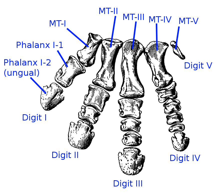
Anterior view of right pes of Centrosaurus; image courtesy of The Open Dinosaur Project
Excellent overview of dinosaur anatomy, and another here, and yet another one here as well.